THE ENDOCRINE SYSTEM: HUMAN HORMONES

There are two types of hormones: steroid & non-steroid. Steroidal hormones are made from cholesterol and are soluble in lipids, meaning they can travel well through a plasma membrane. These hormones target intracellular receptors because of their ability to get through a cell. Steroid hormones are activated once they bind to protein receptors WITHIN a cell. The new hormone-receptor group then binds to DNA to turn a gene on or off. Aldosterone operates this way and creates mRNA. Non-steroidal hormones are made from proteins and are not able to diffuse through a cell membrane, making them more reliant on external receptors outside of the cell. NS hormones rely on a messenger system to create the desired effects of a hormone. The 1st messenger causes the initial reaction outside the cell that then causes cAMP to be released as a 2nd messenger. The reaction moves like a family tree--it starts small and eventually grows with many branches of "output". cAMP works to activate kinases that transfer phosphate groups from ATP to other molecules, which can inactivate or activate them.
The endocrine system has 3 ways of being stimulated: humoral, neural, and hormonal. Humeral stimulation begins with a drop or rise in concentration of materials in the blood (ex. ions and nutrients). Neural stimulation happens with, for example, stress. Stress causes sympathetic fibers in the brain to release Norepinephrine and Epinephrine (from the adrenal gland). Hormonal stimulation is when one hormone affects the activity of another. An example of hormonal stimulation is when the hypothalamus of the brain releases Releasing Hormone that stimulates cells of the anterior pituitary gland.
The endocrine system has 3 ways of being stimulated: humoral, neural, and hormonal. Humeral stimulation begins with a drop or rise in concentration of materials in the blood (ex. ions and nutrients). Neural stimulation happens with, for example, stress. Stress causes sympathetic fibers in the brain to release Norepinephrine and Epinephrine (from the adrenal gland). Hormonal stimulation is when one hormone affects the activity of another. An example of hormonal stimulation is when the hypothalamus of the brain releases Releasing Hormone that stimulates cells of the anterior pituitary gland.
THE PITUITARY GLAND: ANTERIOR & POSTERIOR

The pituitary gland is separated into anterior and posterior regions. The anterior pituitary is glandular and has a hypophyseal "portal" connecting it to the hypothalamus. The anterior part of the pituitary manufactures hormones. Posterior pituitary, in contrast, does not make hormones, it just secretes them. It is neural tissue, meaning hormones are released by synapse rather than vessels. The posterior pituitary is connected to the hypothalamus via the hypophyseal "tract" rather than the "portal".
ANTERIOR PITUITARY HORMONES:
1. Growth hormone (protein): this hormone is made by somatotrophic cells. It encourages the body to use fats for fuel and stimulates cell building. It directly affects elongation of bone and indirectly affects the growth of soft tissues. A hyper-secretion of this hormone causes gigantism if started in childhood and acromegaly if started in adulthood. The treatment for this is removal of the pituitary gland. Click the button below to watch a video about these conditions.
2. Prolactin hormone (protein): Prolactin is made by lactotrophic cells. this hormone is a protein that promotes milk production and is regulated by the hypothalamus. Dopamine inhibits the production of the hormone.
3. Thyroid Stimulating Hormone/Thyrotropin (glycoprotein): Made by thyrotropic cells. TSH stimulates growth of the Thyroid gland and controls secretions of thyroid hormones. With too much TSH, one can have hyperthyroidism. This condition is called Graves disease and results in a goiter, or oversized thyroid (throat area). Hypothyroidism (low T3 & T4) results from Iodine deficiency (because TSH works well only with Iodine) or during pregnancy. This hormone displays negative feedback patterns, as the level of thyroid hormone rises, TSH and TRH levels decrease and thyroid hormones (T3, T4) stop being released.
4. Adrenocorticotropic hormone (peptide): This is made from corticotropic cells in the anterior pituitary gland. ACTH functions to stimulate the cortex of the ADRENALS that secrete glucocorticoids--which regulate a body's sugar use and most importantly, helps maintain biological processes when a person is stressed. It is regulated with CRH (corticotropin-releasing hormone) that is released from the hypothalamus and travels to the adrenal cortex. CRH reduces stress and levels are higher when you wake up in the morning.
5. Follicle Stimulating Hormone (glycoprotein): This is made by gonadotropic cells and targets the gonads. This hormone stimulates the follicles in the ovary to mature an egg. It also stimulates the production of sperm in the testes.
6. Luteinizing Hormone (glycoprotein): This hormone promotes the secretion of sex hormones in BOTH males and females. A "surge" of LH is necessary for a female to release an egg, or ovulate. This hormone peaks at day 12 of the menstrual cycle.
Note: Both FSH & LH are controlled by GnRH (gonadotropin releasing hormone) that is secreted from the hypothalamus, travels to the anterior pituitary, and eventually the gonads.
POSTERIOR PITUITARY HORMONES:
How they work: These secretions are not neurotransmitters. The hormones travel down axons, go through the hypothalamus, through the pituitary stalk, and into the posterior pituitary. They are stored in vesicles & the end of axons here. They are released when a nerve impulse passes through.
1. Anti-diuretic hormone (polypeptide): This is produced by neurons in the hypothalamus. It decreases urine production by making the kidneys reduce the amount of excreted water. It is called "vasopressin" for its effects on blood vessels. ADH increases BP and the secretion of it increases after severe blood loss. It is regulated by osmoreceptors (sense change in fluid concentrations) in the hypothalamus. When you are dehydrated, solutes in your blood are more concentrated. This is sensed and the posterior pituitary releases ADH to keep water in the body.
2. Oxytocin: Oxytocin has ADH properties but not as strong. This hormone causes contraction of smooth muscle (ex. Labor contractions) and releases milk from glands in the breasts. Oxytocin is one of the few hormones that work with a POSITIVE feedback system (Uterus stretches, signals hypothalamus to release oxytocin, causes contraction) OR (Infant suckling, hypothalamus releases oxytocin into posterior pituitary, ejects milk from glands). OT is also called a cuddle hormone and may play a role in arousal and orgasm.
THE THYROID & PARATHYROID GLANDS

The thyroid gland is made of two lateral lobes that are connected by the isthmus. They are made of secretory follicles--cells that produce and secrete hormones that are stored or released in extra-follicular cells.
Follicular cells make:
T3 & T4: These hormones regulate metabolism and determine calories used for BMR (Basal Metabolic Rate). They help growth and development as well as help to mature your nervous system. T3 & T4 are controlled by TSH (Thyroid stimulating hormone) in the anterior pituitary. Follicular cells need Iodine to to make thyroid hormones. Iodine + tyrosine = thyroid hormone.
Parafollicular (extrafollicular) cells make:
Calcitonin: the main function of this hormone is to inhibit osteoclasts (cells that break down bone). It keeps Ca out of the bloodstream and leaves the body with reduced circulating calcium. Calcitonin is stimulated by Ca concentration in the blood. Rises are seen after meals and during pregnancy (to protect mother from losing bone mass).
The Parathyroid Gland is located on the back of the thyroid. There are 4 total, 2 superior and 2 inferior. The parathyroid produces parathyroid hormone (protein). PTH increases blood calcium by stimulating osteoclasts to break down bone and stop osteoblasts from building bone. The aftermath of this is higher blood Ca. PTH increases the amount of Vitamin D in the body and allows more Ca to be absorbed through the intestines and into the blood--raising Ca levels. PTH is the counteract to Calcitonin, both maintain balance of Ca levels in the blood.
THE ADRENAL GLANDS

The Adrenal Glands are associated with the kidneys. Adrenal glands are made up of two parts--the medulla and the cortex.
The Medulla: made up of Chromaffin cells that produce amines called norepinephrine & epinephrine (epinephrine is synthesized from norepinephrine). These are part of the sympathetic nervous system. They increase heart rate, blood pressure, and decrease digestive activity. Medulla hormones last up to 10x longer than neurotransmitters and stay in circulation longer.
The Cortex: this part makes up most of the adrenal gland. It is composed of three layers. Starting with the outermost layer:
1. Zona glomerulosa: release mineralcorticoids (balance water and minerals in blood). Example is aldosterone that reduces Na output through urine. This is a reason for bloating during the menstrual cycle.
2. Zona fasciculate: thick layer, release glucocorticoids (cortisol, stress hormones). Cortisol is made by neogensis (making glucose from fats. Cortisol inhibits CRH & ACTH mentioned above).
3. Zona reticularis: makes estrogen and testosterone. Testosterone influences female sex drive.
THE PANCREAS

The pancreas is responsible for both exocrine functions (digestive juices) and endocrine functions (insulin & glucagon in blood).
Glucagon: stimulates liver to break down glycogen (stored sugars). This raises blood sugar by using gluconeogenesis (from fats). When you have a low blood sugar, glucagon is released from ALPHA cells. Release of glucagon stops when blood sugar is back to normal.
Insulin: This works opposite of Glucagon. Instead raising blood sugar, insulin takes sugars out of the blood, lowering blood sugar. It is released from BETA cells. It stimulates the liver to take glucose and store it as glycogen--moving glucose from blood and into cells. Insulin stops being released when blood sugar is low.
THE PINEAL GLAND
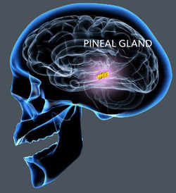
The Pineal Gland is located between the two hemispheres of the brain, near the 3rd ventricle. It is in control melatonin (synthesized from serotonin), which is released depending on light and dark. When low light hits the retina, impulses through the nerve go to the hypothalamus and through the spinal cord, through the sympathetic nerve and then to the brain stimulating the creation of melatonin.
This is the reason we get sleepy at night or in dim lit rooms, the lack of stimulation from light "tells" our brain to make melatonin (which makes us tired!). Less light = more melatonin. This is also part of fertility cycles in some mammals.
THE THYMUS

The Thymus lies in the mediastinum, between the lungs. It is larger in children than it is in adults. Over time, it shrinks and becomes more fatty tissue. The thymus is responsible for secreting thymosins that influence the making of white blood cells (WBC's). It plays a huge role in immunity. It is seen as a "school" for immature WBC's, where they learn to defend the body from potential invaders.
