THE DIGESTIVE SYSTEM - how it all works together
digestive system - The basics
There are two parts that make up the digestive system--the alimentary canal and accessory organs. The alimentary canal is where the food ACTUALLY passes through, whereas the accessory organs contribute without holding the food. The alimentary canal includes the mouth, pharynx, esophagus, stomach, and the small & large intestines. Accessory organs include the teeth, tongue, gallbladder, pancreas, salivary glands, and the liver.

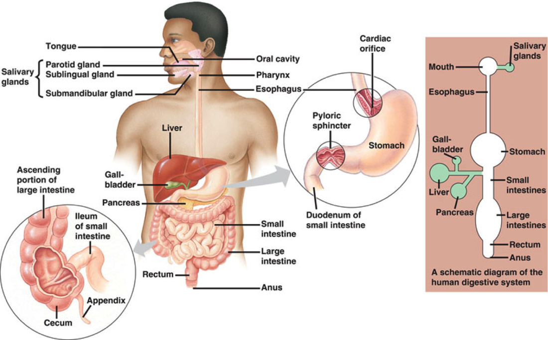
ORGANS & GLANDS OF DIGESTIVE SYSTEM
SO, how does it all work?
The digestive system works by moving food with contractions through the alimentary canal (peristalsis). Most people think that our stomach is our primary digestive organ, but really, that doesn't give enough credit to the small intestine. The stomach works to churn food and chemically digest foods with acids. As the food (chyme) goes through the small intestine, that is where the back and forth motions happen, food breaks up even more, and then absorption occurs! Really, the small intestine does a majority of the work for our digestive system. This is also the location where the gallbladder releases bile to "spread out" fat molecules for absorption and exposure to lipase (from the pancreas). The large intestine does most of the water-absorption, allowing for stool to form and eventually be expelled.
Ever wonder why, after a big meal, you feel very full for a long period of time? In our GI tract, there are receptors that sense stretch, pH, and substrate (fats, proteins). The more substrate consumed, the slower your digestion will be. This is also why you might hear to eat a protein rich breakfast before school, or before a long hike. Protein slows the digestive system down and keeps you fuller, longer!
Besides that, it is interesting to look at the histology of the digestive system to understand a little more how it works and keeps on working. From the esophagus to the anus, the alimentary canal is made of the same 4 layers throughout. From the inside out, there is the mucosa, submucosa, muscularis externa, and the serosa. The space inside of the intestines is called the lumen. The mucosa layer on the innermost portion of the alimentary canal secretes mucus through goblet cells, absorbs through the columnar cells, and is protected with mucosa-associated lymphoid tissue (MALT). The functions are countless! The mucus secreted protects organs from digesting themselves and helps lubricate chyme. In the stomach and small intestine, there are exocrine enzyme (because the alimentary canal is considered an external tube because it is open at both ends) secreting cells that aid in digestion. The submucosa holds lymphatic tissues and elastic fibers. The muscularis externa is the smooth muscle (involuntary) responsible for peristalsis and movement of food down the gastrointestinal canal. The digestive system is more complex than expected, isn't it?
Parts of the Digestive System:
Salivary glands: produce saliva that keeps the mouth clean, as well as contains amylase to start the breakdown of starches. This is why when you eat a piece of bread, it starts to taste sweet. Your saliva is partially digesting the sugars in your mouth!
Pharynx: allows food, fluids, and air through it to go to their respective locations (esophagus or trachea). Lined with stratified squamous epithelium and mucus glands. These help to protect the pharynx from abrasion (lots of food going through).
Esophagus: This is an important place because it moves the chewed food (bolus) into the stomach. It is muscular and contracts similarly to the small intestine with peristalsis. The muscle type changes from skeletal (voluntary) in the superior portion to smooth (involuntary) in the inferior portion. You can say, from knowing this, that swallowing (also called deglutition) uses BOTH voluntary and involuntary muscle of the esophagus. Swallowing uses 22 different muscle groups.
Stomach: This where protein is first broken down. It is smooth muscle that is controlled by temperature, stress, and hormones. If you're in panic mode, digestion will slow and if you are relaxed, it will speed up. The lining of the stomach is made with goblet cells that secrete an alkaline (basic) mucus that helps protect the stomach from its own acid. Special cells make up the stomach:
1. mucous neck cells - acid mucus
2. parietal cells - HCl & intrinsic factor (helps absorb B12). HCl secretion is stimulated by AcH, histamine, and gastrin (causes more chemicals to make HCl)
3. Chief cells - pepsinogen (activated to pepsin by HCl) that breaks down proteins (eggs, meats)
4. Enteroendocrine cells - secrete intestinal hormones
An important thing to note about the stomach is that it has two sphincters--one called the lower esophageal sphincter (LES) and another called the pyloric sphincter. The LES should not be an open gate, or we would get heartburn all the time! However, the pyloric sphincter is supposed to have open/close "gate", so that it can let food in and out to help churn and mix contents.
a. Cephalic phase = (think head), before eating, this is you thinking about food, smelling food (all in your head)
b. Gastric phase = this starts once the food enters your stomach, more and more "juices" are secreted. Gastrin is release during this phase and the stomach stretch receptors are activated. Emotional upset can disturb this phase.
c. Intestinal phase = turn off secretions, once food is sensed in the duodenum. A low ph and duodenal activity starts up the intestinal phase. Fatty chyme slows the intestinal phase.
Small Intestine: made up of the duodenum, jejunum, & ileum.
In the duodenum, bile is released from the gallbladder (gated by sphincter of Oddi) and the pancreas secretes lipase. The duodenum is where all of the secretions take place that help break down and absorb food. Brunners glands in the duodenum secrete an alkaline mucus that helps to neutralize the highly acidic chyme coming from the stomach. Peyer's patches line the intestines (like little police stations) to help maintain "security" and distribute lymph fluid to clean out pathogens in the gut. As you move further down the small intestine, there are less enzymes and there is more water-absorption going on.
Digestion in the small intestine works to digest carbohydrates and proteins, partially. So far, no fat digestion has taken place. Because of the acidic nature of the chyme, the duodenum must receive chyme and push it back to the stomach--back and forth, in order to slowly neutralize the chyme. The small intestine works best at segmentation (stimulated by pacemaker cajal cells) which is the back and forth movement (compared to the one way movement of peristalsis). Towards the end of the small intestine, all of the nutrients have been absorbed. The leftover chyme is turned into a mass of meal remnants, bacteria, and mucosal cells that moves into the large intestine.
Liver: largest GLAND in the body with 4 lobes and it is connected to the gallbladder. It is important to know that the liver CREATES bile and the gallbladder STORES it. Bile is made of bile salts, pigments, fats, electrolytes, and cholesterol. Bile works to "emulsify" or unravel fat chains to expose them to lipase (enzyme that breaks down fat). Bile is released when acidic chyme reaches the duodenum and cholecystokinin (CCK) and secretin into the bloodstream. CCK targets the gallbladder to release bile into the duodenum.

Pancreas: Has both exocrine (outside of body--including GI tract) and endocrine (in body) function. The pancreas, in the GI tract, secretes pancreatic juice and clusters of secretory cells (acini) contain zymogen with digestive enzymes. The endocrine function deals with releasing chemicals (insulin, glucagon) into the blood. The pancreatic juice is mostly a basic HCO3 that helps neutralize acidic chyme.
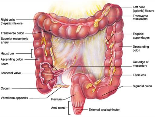
Large Intestine: This part of the intestine is not as active as the small intestine (with all of the exocrine ducts), but there are some fascinating parts of it that help our body to be great at well, digesting! If you look on the image below, on the right side, you can find the label for the "tenia coli". These are bands of longitudinal smooth muscle in the muscularis. The tenia coli help to bind food particles with mucus and create fecal matter along with the haustrum--structures that contract and move food when they sense stretch in the large intestine. Besides these structures, the anatomy of the large intestine also includes the cecum, appendix, colon, rectum, and the anal canal. The most important part of the large intestine is, for sure, the colon. The colon works to, "to accept and stores food remains that were not digested in the small intestine; and to eliminate solid waste (feces) from the body" (7). Bacteria in the colon eat away at fiber and create smooth bowel movements. We have our colons to thank for making fiber a great fighter against colon cancer by helping movement and colon emptying. These bacteria also synthesize vitamins B & K. "Over one hundred trillion microorganisms (bacteria) call the colon home" (7). Simple columnar cells line the colon, with numerous crypts and goblet cells to produce a lubricated movement. Interesting Fact: Without a colon, life would be uncomfortable, but we would survive! It is not needed for life.
**Hemorrhoids happen when the veins of the anal canal are inflammed--they become itchy and uncomfortable.
LARGE INTESTINE
chemical digestion - absorption
Carbohydrates: When carbs are broken down, they then flow into the capillary beds of the villi in the small intestine. They move to the liver via the hepatic portal vein. Salivary amylase, pancreatic amylase, and brush border enzymes help with the carbohydrate digestion process.
Proteins: The absorption of proteins is similar to that of carbohydrates. Pepsin (inactive form: pepsinogen) is an enzyme in the stomach that starts the protein breakdown. Pancreatic enzymes and brush border enzymes further the process.
Fats: These are a little different from the carbs and proteins. For fat to be absorbed, it must dissolve into intestinal cells, combine with protein, and finally move in the lacteals of the microvilli. Carbs and proteins will circulate ONLY in the blood, whereas fats travel through the lymphatic system fluids to reach the blood. They are finally absorbed into capillary blood and transported in the portal vein with help from bile salt [physical digestion] (emulsifier--spreads the fat molecules out to expose them to lipases) and pancreatic lipase [chemical digestion].
Nucleic acids: nucleic acids must be absorbed using active transport, energy (ATP), with membrane carriers. They are absorbed into the villi and moved to the liver through the hepatic vein.
PROBLEMS WITH NUTRIENT ABSORPTION: anything that disrupts absorption can cause malnutrition. Malnutrition could be a reuslt of damage to the intestines, gluten issues (celiac disease--all gluten damages intestines), reduces the length of the microvilli = less absorption. Cancer also has an effect on absorption. Colon cancers usually become liver cancer. The best prevention, as GI cancer rarely shows symptoms, is to have regular dental care and attention to heart health.
The URINARY system
As you know, the systems of the human body are very much connected. The urinary system plays a role in absorption and helps transport nutrients as the digestive system does. The kidneys filter 200 liters of blood daily--they have a lot of pressure to keep our bodies going. Kidney failure is so lethal because without filtration of blood and fluids, toxins build up. The kidneys also help to maintain balance between water, salts, acids, and bases. Gluconeogenesis is taken up by kidneys too, where during fasting, our body can still retrieve a source of carbohydrate. Furthermore, the kidneys make renin (regulate blood pressure) and erythropoietin (stimulate RBC production) which in turn, activates vitamin D. The kidneys are an amazing set of organs and we could not live without them.

Path through the urinary system
The path through the urinary system is as follows:
Bowman's capsule (Glomerular Capsule), Proximal Convoluted Tubule, Descending Limb, Loop of Henle, Ascending Limb, Distal Convoluted tubule, Collecting duct, Papillary Duct, Minor Calyx, Major Calyx, Renal Pelvis, Ureter, Urinary Bladder, & finally the Urethra. It is a long pathway, so if you need more explanation with a visual, watch the video below.
Bowman's capsule (Glomerular Capsule), Proximal Convoluted Tubule, Descending Limb, Loop of Henle, Ascending Limb, Distal Convoluted tubule, Collecting duct, Papillary Duct, Minor Calyx, Major Calyx, Renal Pelvis, Ureter, Urinary Bladder, & finally the Urethra. It is a long pathway, so if you need more explanation with a visual, watch the video below.
An interesting part of the kidney is the nephron. The nephron is the unit that creates urine in the kidney. About 85% of them are located in the cortex of the kidney. When you move closer to the capillary beds, pressure increases, and fluids & solutes are forced out of the blood and into the corpuscle.
The rate of glomerular filtration depends on intrinsic controls, neural controls, and hormonal controls. If GFR is too high, substances needed in the body aren't able to stick around long enough to be absorbed. If GFR is too low, then everything that needs to leave the body will be reabsorbed instead (including wastes). Intrinsic control usually maintains a constant rate. Extrinsic control, however, depends on the activity of the sympathetic nervous system. When the sympathetic nervous system is at rest, kidney blood vessels are dilated and the system auto-regulates itself. When a person is under stress and the sympathetic nervous system is readily active, norepinephrine and epinephrine (from adrenal medulla) are released and the afferent arterioles constrict, inhibiting filtration. The sympathetic system also stimulates activation of the renin-angiotensin mechanism, which starts as soon as juxtaglomerular cells release renin. To watch this play out, refer to the video below this image! It is a great visual.
The rate of glomerular filtration depends on intrinsic controls, neural controls, and hormonal controls. If GFR is too high, substances needed in the body aren't able to stick around long enough to be absorbed. If GFR is too low, then everything that needs to leave the body will be reabsorbed instead (including wastes). Intrinsic control usually maintains a constant rate. Extrinsic control, however, depends on the activity of the sympathetic nervous system. When the sympathetic nervous system is at rest, kidney blood vessels are dilated and the system auto-regulates itself. When a person is under stress and the sympathetic nervous system is readily active, norepinephrine and epinephrine (from adrenal medulla) are released and the afferent arterioles constrict, inhibiting filtration. The sympathetic system also stimulates activation of the renin-angiotensin mechanism, which starts as soon as juxtaglomerular cells release renin. To watch this play out, refer to the video below this image! It is a great visual.

